
What is Abdominal / Transvaginal Ultrasound?
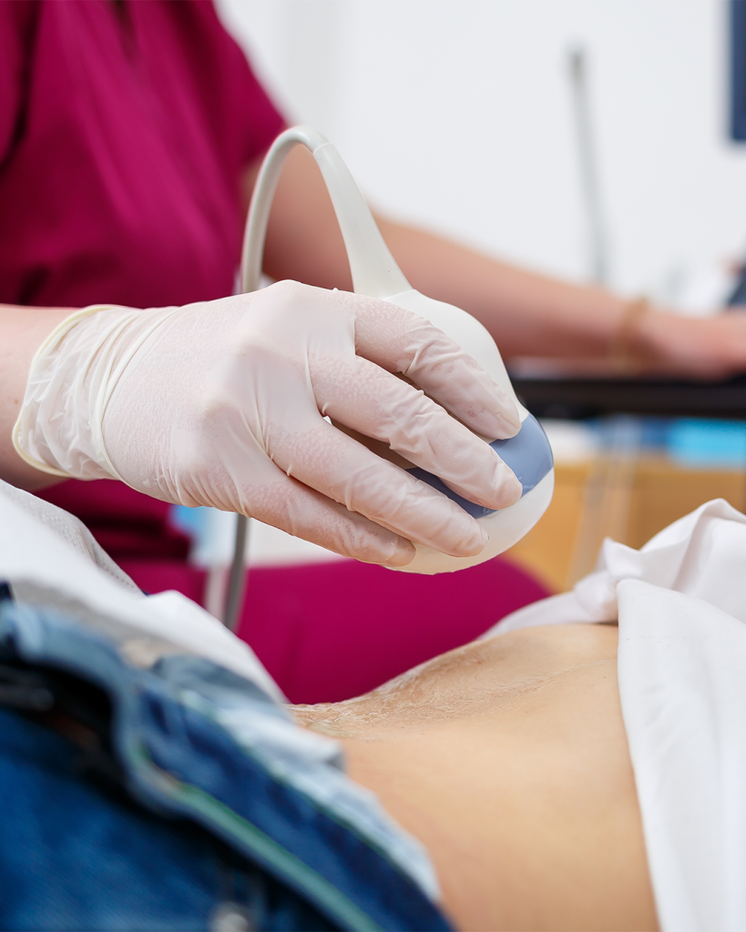
Ultrasound imaging is a non-invasive, essential tool for assessing reproductive and gynaecological health. At EPIA, we use two types of scans: abdominal ultrasound (performed over the abdomen) and transvaginal ultrasound (a closer internal scan using a small probe). These imaging techniques help evaluate the uterus, ovaries, and surrounding pelvic structures safely and effectively.
Abdominal ultrasound is useful for broad anatomical assessments, while transvaginal ultrasound offers more detailed views—especially valuable for fertility evaluations, cycle tracking, and treatment monitoring. Together, they provide real-time insight into follicle development, endometrial lining, and possible abnormalities.
Who is Ultrasound Imaging For?
- Women undergoing fertility evaluations, ovulation tracking, or IVF
- Patients experiencing menstrual irregularities or pelvic pain
- Individuals being monitored during egg freezing, IUI, or IVF cycles
- Women with known or suspected ovarian cysts, fibroids, or uterine concerns
- Anyone needing a baseline reproductive health scan as part of consultation or treatment
The EPIA Difference
Why Choose EPIA for Ultrasound Imaging?
- State-of-the-art ultrasound machines optimised for fertility use
- Scans performed by fertility specialists—not technicians
- Cycle-specific imaging for follicle tracking, ovulation monitoring, and lining assessment
- Immediate scan interpretation with next-step planning
- Comfortable, clean, and patient-sensitive care environment
Transformation
What is the Ultrasound Process?
- Step 1: Appointment Scheduling and Instructions - You’ll be advised whether an abdominal or transvaginal scan is needed and guided on any preparation—such as arriving with a full bladder for abdominal imaging.
- Step 2: Imaging Procedure - The abdominal scan involves moving a probe over your lower belly. For transvaginal ultrasound, a slender probe is gently inserted into the vaginal canal to get a clearer view of the uterus and ovaries.
- Step 3: Visual Assessment - The scan evaluates follicle count and growth, uterine shape, endometrial lining, cysts, fibroids, or any other findings relevant to your cycle or concern.
- Step 4: Doctor Review and Plan - The results are explained to you immediately and incorporated into your fertility or gynaecological care plan on the same day.
Ultrasound
FAQs
Can't find what you're looking for?
What is the difference between abdominal and transvaginal ultrasound?
Abdominal scans give a general view of the pelvic organs and are done over the skin. Transvaginal scans use a slim internal probe to provide a closer, more detailed view of the uterus and ovaries.
Is the scan painful?
Abdominal scans are painless. Transvaginal scans may cause slight pressure but are generally quick and well tolerated.
When is an ultrasound typically done?
For fertility purposes, ultrasounds are timed during the early or mid-cycle to assess follicles, ovulation, and the thickness of the uterine lining.
What conditions can be detected with ultrasound?
Ultrasounds can help identify ovarian cysts, fibroids, endometrial polyps, uterine septum, fluid retention, and track follicular or endometrial changes during treatment.
Do I need to prepare before the scan?
For abdominal scans, a full bladder is recommended. Transvaginal scans do not require any special preparation.
How long does the scan take?
Typically 10 to 15 minutes. Results are shared during the same visit.
Are these scans safe?
Yes. Ultrasound uses sound waves—not radiation—and is completely safe for all diagnostic and treatment-related fertility care.